Background
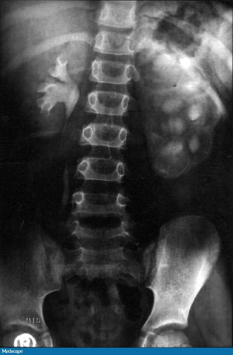 Figure 1. (Click to enlarge) |
A 9-year-old boy is brought to the outpatient clinic by his parents with a 2-3 year history of intermittent fever, abdominal pain, failure to thrive, tiredness, and weakness. In between times of active illness, the patient is usually asymptomatic, although he gets tired very easily during his sporting activities. When present, the fever and pain usually subside on their own but, occasionally, an antipyretic or a course of antibiotics is needed. His weight has been constant and there has been no significant height gain over the last year. He does not have any nausea, vomiting, diarrhea, constipation, bloating, or distention. There is no history of hematuria or stone disease. He does not have any past history of surgery, prolonged fever, hospitalization, or any illnesses (except for the present one). There is no significant family history. He has no known allergies, and his vaccinations are up to date. The patient comes from a poor socioeconomic background.
The child is well-nourished but appears small for his age. He is friendly and cooperative. His blood pressure is 94/68 mm Hg, pulse is 80 bpm with a regular rhythm, oral temperature is 98.8°F (37.1°C), and respiration rate is 16 breaths/min. The patient's conjunctiva is pale, the sclera is muddy, the neck veins are not distended, and no pedal edema is noted. Although the abdomen is slightly distended, no organs or lumps are palpable, and the bowel sounds are normal. Slight tenderness is noted in the left lumbar region on deep palpation, while the rest of the abdomen is nontender. The review of his other systems is normal. No free fluid is detected in the abdomen. The general examination does not show any abnormalities, except for slight pallor and grade 1 digital clubbing. No lymphadenopathy is detected, and the hernial sites are normal.
A complete blood cell (CBC) count reveals a hemoglobin of 10.7 g/dL (107 g/L), a hematocrit of 32% (0.32), and a red blood cell (RBC) count of 3.5 × 10⁶/µL (3.5 × 10¹²/L). The liver function tests, kidney function tests, electrolyte panel, and coagulation profile, as well as the remainder of the CBC, are all normal. A routine urine examination shows 3-4 white blood cells (WBCs) per high power field, with no RBCs. A urine culture is performed; within 48 hours, over 100,000 colony forming units per milliliter (CFU/mL) of Escherichia coli sensitive to ciprofloxacin, gatifloxacin, gentamicin, and amikacin are found.
An intravenous urogram is ordered (see Figure 1).
Hint: Compare both the right and left pelvicalyceal systems.
d. Ureterovesical junction obstruction
Discussion
| Figure 1. (Click to enlarge) | 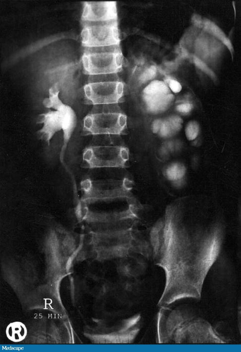 Figure 2. (Click to enlarge) |
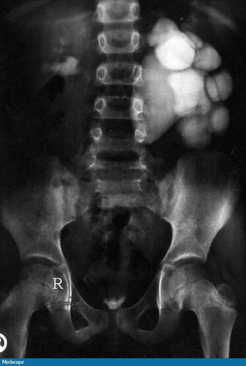 Figure 3. (Click to enlarge) | |
On the initial nephrogram images, a large amount of uptake was noted in the right kidney (this is known as a "prompt nephrogram"); however, on the left side, there was a delay of uptake into the kidney parenchyma (known as a "delayed nephrogram"). (Please note that the initial film obtained in the first minute of the nephrogram is not available.) A delayed nephrogram is pathognomonic for an obstruction. It does not, however, show exactly where the obstruction is located. Figure 1, a 5-minute delayed film, demonstrated a persistent nephrogram in an enlarged left kidney and delayed excretion of contrast into a dilated collecting system; the right kidney appeared normal. In addition, on other delayed films (see Figures 2 & 3), a massively dilated pelvicalyceal system with blunting of the calyces was noted on the left. A cut-off point at the ureteropelvic junction (UPJ) was also noted on that side, without visualization of the left ureter. All of these changes were consistent with localization of the obstruction to the UPJ on the affected side. The left ureter was not seen to opacify on the delayed films. The right pelvicalyceal system, however, was well-defined, and the calyces showed normal sharp cupping (see Figure 2). The right ureter appeared thin or delicate, and a column of contrast was noted to reach down to the bladder; this ruled out any right-sided obstruction (Figure 2). The postvoiding film showed almost complete emptying of the bladder. By that point, the contrast had relatively cleared (with some residual contrast still in the collecting system) from the right kidney collecting system; the left side, however, was still full of contrast (see Figure 3). These findings were consistent for a radiographically established UPJ obstruction.
The ultrasonography (images not available) in this patient showed dilatation of the left pelvicalyceal system, with hydronephrosis of the left kidney and thinning of the cortex. The right kidney and bladder were normal. No abnormalities were seen in the liver, gall bladder, pancreas, or spleen. A renal scan was also performed (images not available), which showed a nonobstructed right kidney with normal function. The left kidney showed hydronephrosis and impaired function suggestive of UPJ obstruction. The total glomerular filtration rate (GFR) was measured at 123.35 mL/min, with a right-kidney GFR of 69.33 mL/min (within normal limits) and a left-kidney GFR of 54.02 mL/min (below normal). The split functions of the kidneys were noted to be 56% for the right kidney and 44% for the left kidney.
UPJ obstruction is the most common obstructive lesion in childhood. Prior to the use of prenatal ultrasound, most patients with UPJ obstruction presented with pain, hematuria, urosepsis, failure to thrive, or a palpable mass. Approximately 60% of cases occur on the left side, and the male-to-female ratio is 2:1. In 10% of cases, UPJ obstruction is bilateral. In kidneys with UPJ obstruction, renal function may be significantly impaired as a result of pressure atrophy.[1,4,5]
Congenital UPJ obstruction most often results from intrinsic disease. A common defect in congenital UPJ obstruction is an aperistaltic segment of the ureter. In these cases, histopathologic studies have revealed that the spiral musculature normally present has been replaced by abnormal longitudinal muscle bundles or fibrous tissue. This results in failure to develop a normal peristaltic wave for the propagation of urine from the renal pelvis to the ureter. Further investigations into the etiology of UPJ obstruction have shown decreased interstitial cells of Cajal at the UPJ in children. In addition, the cytokine produced in the urothelium is also theorized to exacerbate UPJ obstruction. A less common intrinsic cause of congenital UPJ obstruction is true ureteral stricture. True congenital ureteral strictures are most often found at the UPJ, although they may be situated at sites anywhere along the ureter. Abnormalities of the ureteral musculature have been implicated because of the deposition of too much collagen at the site of the stricture. Intrinsic obstruction at the UPJ may also result from kinks or valves produced by infoldings of the ureteral mucosa and musculature. In these cases, the obstruction may be, essentially, at the level of the proximal ureter. Grossly, this can manifest as external bands or adhesions that appear to cause the obstruction. In the majority of cases, however, these bands or adhesions are likely to be a secondary phenomenon associated with intrinsic obstruction; therefore, operative pyeloplasty would generally be the most effective procedure.[1,4,5,6]
The presence of these kinks, valves, bands, or adhesions may also produce angulation of the ureter at the lower margin of the renal pelvis in such a manner that, as the pelvis dilates anteriorly and inferiorly, the ureteral insertion is carried further proximally. In these cases, the most dependent portion of the pelvis is inadequately drained, and the apparent "high insertion" of the ureteral ostium is actually a secondary phenomenon. In at least some cases, however, the high insertion itself is likely the primary obstructing lesion, because this phenomenon is frequently found in cases of renal ectopia or fusion anomalies. As such, a high insertion can have implications in the ensuing surgical management. An accessory artery and/or vein to the lower pole of the kidney also may cause extrinsic obstruction. Although these lower-pole arteries have often been referred to as aberrant, this variant is found in 20% of people. In some patients, these lower-pole vessels cross the ureter posteriorly and truly have an aberrant course. Although some have speculated that the crossing vessel plays a role in the pathogenesis of UPJ obstruction, it is unlikely that the associated vessel alone causes the primary obstruction. In fact, the true etiology is an intrinsic lesion at the UPJ or proximal ureter that causes dilatation and ballooning of the renal pelvis over the polar or aberrant vessel.[2,5,6]
UPJ obstruction may also result from acquired lesions. In children, vesicoureteral reflux can lead to upper tract dilatation, with subsequent elongation, tortuosity, and kinking of the ureter. Other acquired causes of obstruction at the UPJ include benign tumors (such as fibroepithelial polyps), urothelial malignancy, stone disease, and postinflammatory or postoperative scarring or ischemia.[4,6]
UPJ obstruction, although most often a congenital problem, can present clinically at any time of life. In older children or adults, intermittent abdominal or flank pain is a frequent presenting symptom. This is occasionally associated with nausea or vomiting as well. Hematuria, either spontaneous or associated with otherwise relatively minor trauma, may also be an initial symptom. Laboratory findings of microhematuria, pyuria, or frank urinary tract infection might also bring an otherwise asymptomatic patient to the health care system. Rarely, hypertension may be a presenting finding.[2,6]
Radiographic studies should be performed with the goal of determining both the anatomic site and the functional significance of an apparent obstruction. Excretory urography remains a reasonable first-line option for radiographic diagnosis. Classically, findings on the affected side include delay in function associated with a dilated pelvicalyceal symptom. Ultrasonography also has an important role in diagnosing UPJ obstruction. It can be employed in patients whose poor renal function precludes the use of intravenous (IV) contrast studies. Because of its current widespread use, computed tomography (CT) scanning more frequently raises the suspicion of UPJ obstruction. It is best for the CT scan to first be performed without contrast in order to rule out obstructing stones, followed by a scan with contrast to assess for lack of drainage from the pelvis as well as detect any potential tumors in the collecting system (ureter or pelvis). Nuclear renography can also be performed for all patients with suspected UPJ obstruction. Specifically, a technetium 99m–labeled mercaptoacetyltriglycine (MAG3) scan can differentiate the split functions of both the kidneys and can quantify the drainage from each kidney. Specifically, it measures the t , which is the time it takes for half of the radiolabeled MAG3 to wash out of the renal pelvis. In cases of obstruction, the t is typically greater than 15 minutes.[4,5,6]
The following entities should be considered in the differential diagnosis: (1) megacalycosis, a congenital nonobstructive dilatation of the calyces without pelvic or ureteric dilatation; (2) vesicoureteral reflux, with marked dilatation and kinking of the ureter; and (3) midureteral or distal ureteral obstruction (when the ureter is not well visualized on the urogram).[4]
The patient was admitted to the hospital and an open dismembered pyeloplasty was planned. The short obstructing segment of the ureter was resected, and the free end of the ureter was sutured to the open renal pelvis. This anastomosis was performed using 4-0 vicryl. A stent bridging the anastomosis was placed from the kidney to the bladder, and a drain in the perinephric space was inserted. The drain was removed on the sixth postoperative day, and the sutures were removed on the tenth day. The internal stent is typically removed in postoperative week 4-6 with an office-based cystoscopy. The postoperative course was uneventful. While retrograde endoscopic approaches have been described, laparoscopic pyeloplasty is rapidly becoming the standard of care. This minimally invasive approach offers the advantages of less pain, a reduced need for pain medications, a shorter hospital stay, quicker convalescence, and better cosmesis. Most importantly, the results of establishing good drainage from the pelvis with laparoscopic pyeloplasty have been shown to be equivalent to those of open pyeloplasty. The patient was doing well after discharge, and he had gained height and weight at both 3-months' and 6-months' follow-up.[4]
a. Delayed nephrogram (or uptake of contrast by the kidney)
b. Hydronephrosis
c. Lack of peristalsis
d. Lack of contrast in the ureter on drainage films

No comments:
Post a Comment